Diagnostic procedures and health programs
At this modern centre for treatment and recovery, prevention and recreation, apart from the sulphuric thermo-mineral water, healthful mud – peloid and beneficial fresh air, a variety of therapeutic procedures are applied, procedures that are made possible by modern physical medicine and rehabilitation.
There is also a modern laboratory available to our patients and all others who want to check their overall health, and in a short period of time perform various blood and urine analyses, and check their hormonal status and tumor markers.
Lectures by medical staff of our Centre are regularly held and they are aimed for the patients and guests of Specialized Rehabilitation Hospital Banja Koviljaca.
There are also consultations given by professors of Medical School with the University of Belgrade (rheumatologist, orthopedist, physiatrist, pediatric surgeon-orthopedist, internist and angiologist).
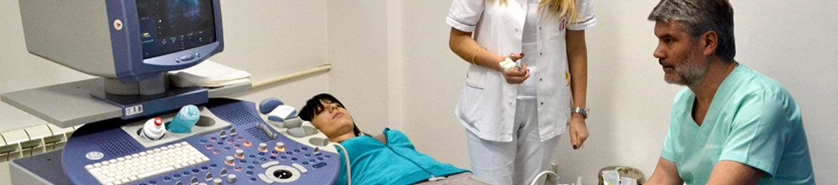
Color Doppler Ultrasound
In Specialized Rehabilitation Hospital Banja Koviljaca you may have Color Doppler Ultrasound of blood vessels. It is non-invasive diagnostic procedure for examination of blood vessels by using ultrasound, where by measuring blood flow velocity and by viewing the inner walls of blood vessels, blood vessel narrowing and expansion may be assessed, and changes in the blood vessel structure and length of the vessel may all be viewed. In comparison to other diagnostic procedures, Color Doppler Ultrasound has advantage for not being invasive, as well as for its high sensitivity and specificity of examination.
With Color Doppler Ultrasound, blood vessels in the neck, arms, legs, abdominal aorta and visceral branch may be viewed.
Neck blood vessels Doppler implies viewing of carotid arteries (common carotid arteries, extracranial segment of outer and inner carotid artery) as well as viewing of vertebral arteries.
With this examination, some changes in arteries may be viewed: narrowing, expansion, tortuosity. If these changes are considerable, then further diagnostics is recommended and a treatment provided by a vascular surgeon. In some less pronounced changes, it is recommended to have monitoring of the changes in six months or in a year.
Indications for examination of carotid tree are the following: permanent headaches, dizziness, blurred vision, weakness of one body part, numbing of one face half, double vision, noise and pulsation mass on the neck, transient ischemic attack (TIA), amaurosis fugax, reversible ischemic neurological deficit ( RIND), vascular dementia, hypertensive encephalopathy, conditions after surgery of carotid artery tree.
Color Doppler Ultrasound is recommended to everybody older than 65.
Color Doppler Ultrasound of legs implies examination of veins (surface, deep, communicating) as well as of arteries. In veins, signs of expansion, blood flow and blood clots within inner or surface veins may all be viewed. In arteries, artery wall, narrowing (presence of plaques), clogging as well as aneurism may all be viewed.
Indications for leg veins examination mostly include a sense of heaviness in a leg (most often lower leg), swelling accompanied with redness, increased skin temperature. Indications for leg arteries examination mostly include a sense of cold in legs, pain and cramps, most often occurring during walking.
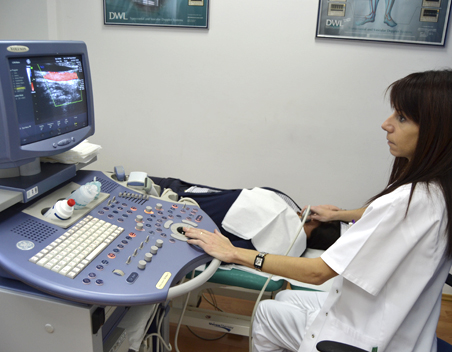
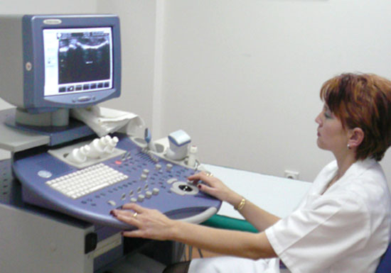
Ultrasonography
Significance of ultrasonography in diagnostic of rheumatoid arthritis
Development of ultrasonography for the past 10 years has enabled use of ultrasonography in diagnostics for musculoskeletal system. The advantage of ultrasonography in comparison to CT, MRI and X-rays is complete absence of ionizing radiation, relatively low price of examination and broad availability of the device. Ultrasonography can differentiate pathological changes in muscles, joints. Ultrasonography helps in making early diagnosis of rheumatic diseases, differential diagnosis for some conditions, and monitoring of the disease course.
It is not possible to view the inner structure of a bone, but the bone cortex and cartilage can be viewed. Foreign bodies are hyperechogenic and these can be viewed best when located within hypoechogenic tissues such as muscles. Hematoma in muscles occurred due to injury can be viewed and in this way localization of hematoma is possible.
For the past 10-15 years, ultrasonography has become an important method for examination and monitoring of patients suffering from RA because it is broadly available, enables viewing of inflammatory changes in synovia, joint liquors, changes in paraarticualr structures.
Bone densitometry – DXA (Bone density measurements)
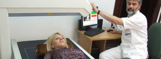
Bone densitometry is a diagnostic method for measuring bone mineral density, i.e. bone strength and it is the only valid procedure either for excluding or final confirmation of osteoporosis.
Significance of early diagnostic is in prevention of fractures, and with timely detection of the disease and initiation of adequate treatment, bone fractures may be prevented and these are the greatest hazard and complication of osteoporosis.
In everyday practice, as per WHO gold standard for diagnostic of osteoporosis, bone density measurements are done on lower back and hip.
In addition to this, the equipment software may calculate a fracture risk, i.e. a risk for developing of great bone fracture and as a special parameter, a risk for developing hip fracture as the most dangerous fracture.
The equipment also enables measuring of complete bone mass, i.e. strength of the complete skeleton in all bones and assessment of structural composition i.e. percentage of bone and muscle mass and fat tissue percentage, being of particular significance, and such finding may be used for management of overweight, i.e. in monitoring body weight reduction and functional recovery of sportsmen in order to make assessment for returning to their training activities.
Our equipment is modern version of Lunar X-ray central scan intended just for bone densitometry of the spine and hip. Scanning is reliable and it is not harmful. The only counterindication is pregnancy, and it does not require any particular preparation or protection. It also involves interpretation of findings, i.e. recommendations for management of osteoporosis if it is diagnosed or advices and instruction for prevention measures in order to prevent its development and all complications followed after it.
ENMG - Electromyoneurography
Electromyoneurography is a diagnostics method for establishing illness and damage of peripheral nervous system and muscles. It implies testing of a part of nervous system from a nerve cell body in spinal cord (motor neuron), through nerve roots (radix), plexus and peripheral nerves, neuromuscular junction (synapsis) and muscles.
The most common conditions where this diagnostic procedure should be done include:
- degenerative disc disorders (herniated disk) in order to find whether disc makes compression onto the nerve roots and their consequent damage,
- injuries of arm nerves during childbirth for establishing level and degree of damage, providing prognosis and planning of therapy program,
- all traumas where there are clinical signs of peripheral nerve injury
- metabolic diseases, such as diabetes mellitus in order to find whether complications of the primary disease have developed, such as diabetic polyneuropathy
- all neurological conditions which affect peripheral nervous system (motor neuron disease, synapsis disease, neuropathies) and muscle conditions (myopathies, dystrophies, and similar).
Therefore, this method is not for examination of central nervous system.
In our Hospital, EMNG examination may be done every work day, with previous appointment made via phone +381 15 895 273.
Our expert consultant is Prof. Dr Sci. Med. Vedrana Milić-Rašić, a specialist in neuropsychiatry, from Child and Adolescent Neurology and Psychiatry Clinic in Belgrade.
Laboratory
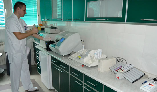
The up-to-date equipped laboratory in Specialized Rehabilitation Hospital has top equipment and many analysis can be done. Patients, as well as all interested clients who would like to check their health status, may have many blood and urine analysis, hormone status and tumor marker in quite short time.
The unique treatment for children in the world
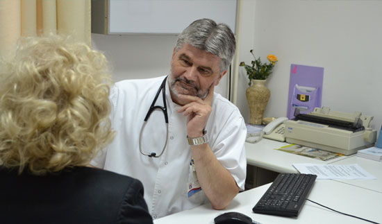
There is a possibility to have consultative examination by Belgrade Medical School professors (rheumatologist, orthopedist, physiatrist, children’s surgeon, internist, angiologist). More on consultants and appointing here.